
2003 Nobel Prize
Reflecting the fundamental importance and applicability of MRI in medicine, Paul Lauterbur of the University of Illinois at Urbana-Champaign and Sir Peter Mansfield of the University of Nottingham were awarded the 2003 Nobel Prize in Physiology or Medicine for their “discoveries concerning magnetic resonance imaging”. The Nobel citation acknowledged Lauterbur’s insight of using magnetic field gradients to determine spatial localization, a discovery that allowed rapid acquisition of 2D images. Mansfield was credited with introducing the mathematical formalism and developing techniques for efficient gradient utilization and fast imaging. The actual research that won the prize was done almost 30 years before, while Paul Lauterbur was at Stony Brook University in New York.[citation needed]
The award was vigorously protested by Raymond Vahan Damadian, founder of FONAR Corporation, who claimed that he invented the MRI, and that Lauterbur and Mansfield had merely refined the technology. A group called “The Friends of Raymond Damadian” (formed by Damadian’s company, FONAR[42]), took out full-page advertisements in the New York Times and The Washington Post entitled “The Shameful Wrong That Must Be Righted”, demanding that he be awarded at least a share of the Nobel Prize.[43]
To perform a study the patient is positioned within an MRI scanner which forms a strong magnetic field around the area to be imaged. Most medical applications rely on detecting a radio frequency signal emitted by excited hydrogenatoms in the body (present in any tissue containing water molecules) using energy from an oscillating magnetic fieldapplied at the appropriate resonant frequency. The orientation of the image is controlled by varying the main magnetic field using gradient coils. As these coils are rapidly switched on and off they create the characteristic repetitive noises of an MRI scan. The contrast between different tissues is determined by rate at which excited atoms return to theequilibrium state. Exogenous contrast agents may be given intravenously, orally or intra-articularly.[17]
MRI requires a magnetic field that is both strong and uniform. The field strength of the magnet is measured in tesla - and while the majority of systems operate at 1.5T commercial systems are available between 0.2T – 7T. Most clinical magnets are superconducting which requires liquid helium. The lower field strengths can be achieved with permanent magnets, which are often used in “open” MRI scanners for claustrophobic patients.[18]
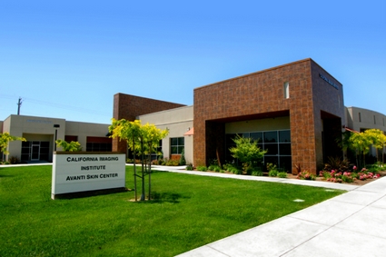
California Imaging Institute is the Central Valley’s newest and most technologically sophisticated outpatient imaging center, offering 3.0T HD MRI, 1.5T Open Bore MRI, CT, Ultrasound, PET-CT, DEXA, Digital X-Ray, and Vascular and Interventional Radiology services.


Share With Friends